Just like the mobile phone, digital camera, and home computer, veterinary radiology technology has evolved over the years. The great thing about this is that as technology evolves, it becomes more widely accessible and offers more and more ways to help us in our daily lives.
Today, we’ll discuss some of the advancements in veterinary fluoroscopy technology, and how these live x-ray studies can help veterinary general practitioners offer new services and provide excellent patient care.
What’s New with Veterinary Fluoroscopy?
Fluoroscopy is a type of live x-ray, performed in real time. Compared to standard radiographs, this provides a more dynamic, functional view of certain parts of the body.
In previous years, a C-arm fluoroscopy unit was the main option available to veterinarians. This type of equipment has been valuable for veterinary patients that needed it but are impractical for all but the largest veterinary practices. However, recent advancements in live x-rays have made fluoroscopy more affordable and more convenient to own and operate.
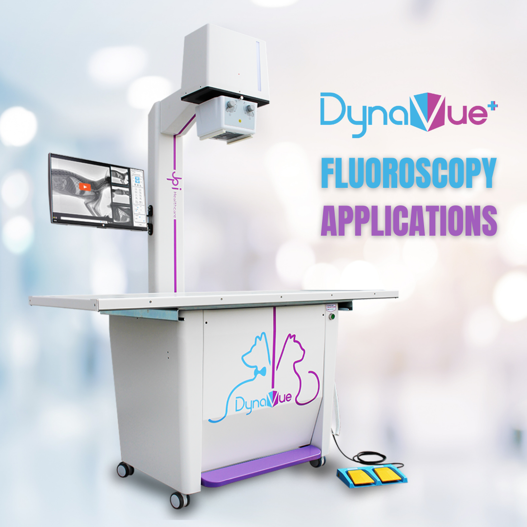
Newer technology includes a 2-in-1 (x-ray system with fluoroscopy) combination system. This offers the convenience of fluoroscopy capabilities without needing to make room in the clinic for a separate large C-arm system, and the ability to seamlessly switch between standard and live x-rays.
Diagnostic Veterinary Fluoroscopy Procedures
For many general practitioners, referral has been the only option for patients that could benefit from a fluoroscopy study. Common examples include diagnosing tracheal collapse and performing a swallowing study.
Some of these patients require ongoing care with a specialist. For example, the most severe cases of tracheal collapse could require a tracheal stent placed by a surgeon who is experienced with that procedure.
However, there are certainly cases where the patient could continue their ongoing care with their regular veterinarian. In these cases, if the practice has fluoroscopy available, keeping the patient in-house becomes an option.
Another example of when live x-rays are valuable is for different types of studies of the urinary tract. This could include moving the patient around in real time to get bladder sludge moving and differentiate it from a mural mass within the urinary bladder. Or, it could include retrohydropropulsion of urethral stones—watching in real time to ensure the uroliths have cleared the urethra and fully made their way into the bladder.
Other exciting examples include live x-ray studies to evaluate for joint instability or laxity due to soft tissue damage (including CCL injuries), GI transit studies, nasopharyngeal polyps, or small fractures within a complex joint such as the carpus or tarsus. While many of these conditions can be diagnosed using standard radiographs, vets may find it’s simpler and faster to see how everything moves in real time.
Real-time views can also make skull and jaw studies easier (and faster), since the required views can be obtained without having to perfectly tape the patient into position each time.
Evaluating for traumatic internal injuries—such as a ruptured urinary bladder diagnosed via a contrast study, or diagnosing a dynamic hernia by watching the movement in real time—is another fascinating application of fluoroscopy in general practice.
Surgical or Therapeutic Procedures Using Veterinary Fluoroscopy
Live x-rays allow for real-time images to aid a veterinarian in certain non-invasive surgical procedures.
Surgical applications will vary from practice to practice, depending on the comfort level of the general practitioner and on the availability of nearby specialists for referral. Examples include non-invasive orthopedic surgeries (such as external skeletal fixators) or closed reduction of a hip luxation.
Fluoroscopy could also aid in procedures such as placement of an esophageal or gastric feeding tube or obtaining guided aspirates/biopsies.
Will Veterinary Fluoroscopy Replace Standard Radiographs or Ultrasound?
While this isn’t a comprehensive list of diagnostic or therapeutic procedures, it covers some of the most common uses. The list of applications is growing as veterinarians who have adopted fluoroscopy into their hospital discover more uses in their daily practice.
Veterinary fluoroscopy isn’t meant to replace standard radiographs or other imaging modalities such as ultrasound. Instead, it can help complete the picture, especially in scenarios such as the ones described above.
There are some health conditions for which fluoroscopy is the ideal method of diagnosis. And in many other cases, vets could find that live x-ray studies give them a new view into what’s going on inside the patient.
These dynamic studies give vets an additional tool, especially when it comes to diagnosing things that happen quickly inside the body—things that could be missed on standard radiographs, which only capture a moment in time. Fluoroscopy also helps with more quickly evaluating parts of the body that would otherwise require multiple standard x-ray views (for example, jaw/skull studies or stress views of joints).
Live x-rays are a helpful tool that provide a unique value compared to other imaging modalities, and they can aid in many diagnostic and therapeutic procedures.
